
Heart diseases are common in pets and often they can be successfully managed or even cured. Whether it is a congenital defect (present from birth) or an acquired heart disease, we are here to help you and your pet.
Our VSH Cardiology Team will make sure that you and your pet are experiencing state-of-the-art care. We offer echocardiographic, radiographic, electrocardiographic (ECG), 24-hour ambulatory ECG evaluations, as well as advanced imaging (angiography, CT). Most patients can be evaluated utilizing non-invasive diagnostic techniques with results readily available. Besides medical management of heart diseases, we also offer minimally invasive interventional procedures to cure or improve certain congenital defects, and pacemaker implantation in patients with abnormal heart rates.
DIAGNOSTIC TESTS
Blood Pressure Measurement
As in people, high blood pressure (hypertension) can have severe health implications. In pets, high blood pressure is usually not a result of heart disease, but can negatively affect the heart as much as the eyes, kidneys, and brain. Common primary diseases that can cause hypertension include kidney disease, hyperthyroidism, Cushing’s disease, Diabetes mellitus, Acromegaly, and Pheochromocytoma. Certain heart diseases, however, can cause a very low blood pressure (hypotension). The blood pressure gives us valuable information that will help us to understand your pet’s condition better and whether the abnormality needs further action.
We measure your pet’s blood pressure in a similar fashion to how it’s done in people. An inflatable cuff is fitted around your pet’s foreleg or foot. With an ultrasound probe, a superficial artery and the pulse of your pet is found. We then inflate the cuff until the Doppler signal of the pulse disappears. The cuff will then be deflated and the Doppler signal returns. This is your pet’s systolic blood pressure, which should not exceed 160mmHg. In order to get a good average reading we will repeat this multiple times. It also allows your pet to get used to the procedure and relax over time.
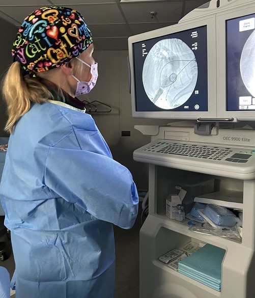
Echocardiogram
An echocardiogram is an ultrasound of the heart. It allows us to evaluate the heart, including quantification of wall thickness, chamber size, valve function, blood flow, and contractility.
The heart is a muscular organ which is located in the chest. The heart functions as a mechanical pump and moves blood through the vessels in the body. The heart has two sides; a right side and a left side. Each side has an upper chamber (atrium) and a lower chamber (ventricle).
Oxygen-poor blood from the body enters the heart at the right atrium, which contracts and moves blood through the tricuspid valve into the right ventricle. The contraction of the right ventricle now pumps blood into the lungs. In the lungs this blood is replenished with oxygen.
The oxygenated blood then returns to the left side of the heart. It is entering first the left atrium. From here the blood is forced through the mitral valve and into the left ventricle. The left ventricle then contracts and pumps blood into the aorta, delivering oxygen-rich blood to the body.
Echocardiography is a non-invasive and low-stress diagnostic test that will be performed with your pet lying on a padded table. In order to get better contact with the ultrasound probe to the skin we will recommend to have a little area around the axillary region clipped of fur. If you would prefer to have your pet not clipped please let us know in advance and we will try to accommodate your wishes. In most cases no sedation (tranquillizers) is necessary. However, some of our patients will be more relaxed if used; we may ask for your permission to do so, if we feel it is in the best interest of your pet.
This test, when performed by an experienced clinician, is essential in diagnosing most heart diseases. Whether it is a disease affecting the heart valves (chronic degenerative valve disease), thickening of the heart muscle (hypertrophic cardiomyopathy), dilation of the heart (dilated cardiomyopathy), congenital heart diseases (patent ductus arteriosus, pulmonic stenosis, subaortic stenosis), or tumors of the heart.
ECG/EKG
The electrocardiogram (also known as ECG or EKG) is a non-invasive test evaluating the electrical activity of the heart. It is performed with your pet lying on the right side with electrodes attached on your pet’s skin. The EKG will help detecting abnormal heart rates and rhythms (arrhythmias). The morphology of the EKG provides also information regarding chamber enlargement and abnormal conduction.
In a normal heart the heartbeat is initiated by the sinus node. The sinus node is a pacemaker region which is located high in the right atrium (link to heart anatomy under echocardiogram). The electrical impulse generated by the sinus node travels through the atria, which as a result then contract. The impulse then passes through specialized cardiac conduction tissue, which rapidly distributes the electrical impulse to the left and right ventricles. The ventricles then contract, and blood is forced blood to the body and lungs. This pattern of electrical impulse production and conduction is repeated for every beat. If at any point the normal conduction is disturbed it will lead to an abnormally looking EKG and allows us to diagnose your pet’s arrhythmia.
Holter monitor
A Holter monitor is a small, wearable device that records the heart’s electrical rhythm for a 24-hour period. The Holter monitor will be fitted to your pet. Different from an in-hospital run EKG, it provides us with a heart rate and rhythm over a much longer period of time. It also allows us to determine the heart rate and rhythm in the home environment, rather than in the veterinary hospital where stress and excitement may confound our interpretation. You will be instructed to take notes on a Holter diary. These are very important information as it helps us to interpret whether the heart rate we are seeing is appropriate or inappropriate (sleep vs. activity). After the 24hrs you may remove the Holter and drop it off at VSH. If you don’t feel comfortable removing the Holter yourself one of our staff members is happy to give you a hand.
Thoracic Radiographs
Radiographs, also known as x-rays, of the chest allow us to assess the heart, lungs, and adjacent structures. This test helps us to diagnose your pet’s heart disease and severity. It is the test of choice to diagnose left-sided congestive heart failure (fluid within the lung tissue) and will also show us the presence of fluid within the chest cavity. This test takes only minutes and can be mostly performed without any sedation (tranquillizers) and only gentle restraint.
Laboratory tests
There are a variety of heart diseases that will lead to an increase in certain markers found in the blood stream (NT-proBNP, Troponin I). Besides these heart-specific markers blood and urine tests give us a lot of valuable information about your pet’s blood cells, organ function, and electrolyte balance. This is not only crucial in order to monitor your pet while on heart failure medication. Many of our patients are older pets; comorbidities are much more common in that population and may need to be addressed as well.
Our Team

Dr. Joel Silva
Specialist in Veterinary Cardiology
Rotating Internship Director
LMV, DipECVIM-CA (Cardiology), MRCVS, RCVS, EBVS
Hong Kong Registered Specialist in Veterinary Cardiology

Dr. Maxie U Krueger
Specialist in Veterinary Cardiology
Dr. Med Vet., DACVIM (Cardiology)
Hong Kong Registered Specialist in Veterinary Cardiology